
FIGURE
Fig. 1
- ID
- ZDB-FIG-191112-1
- Publication
- Akerberg et al., 2019 - Deep learning enables automated volumetric assessments of cardiac function in zebrafish
- Other Figures
- All Figure Page
- Back to All Figure Page
Fig. 1
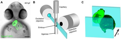
In vivo cardiac imaging with light-sheet fluorescence microscopy (LSFM). (A) Fluorescent image of a 48 hpf Tg(myl7:GFP) zebrafish embryo with bright-field overlay. Scale bar: 100 μm. (B) Schematic depicting the imaging of a live zebrafish embryo immobilized in agarose within the excitation and detection axis of a light-sheet microscope. (C) Schematic of dynamic image acquisition through z-depths. |
Expression Data
Expression Detail
Antibody Labeling
Phenotype Data
Phenotype Detail
Acknowledgments
This image is the copyrighted work of the attributed author or publisher, and
ZFIN has permission only to display this image to its users.
Additional permissions should be obtained from the applicable author or publisher of the image.
Full text @ Dis. Model. Mech.