
Fig. 8
- ID
- ZDB-FIG-191021-17
- Publication
- Lee et al., 2019 - A thrombomodulin-like gene is crucial to the collective migration of epibolic blastomeres during germ layer formation and organogenesis in zebrafish
- Other Figures
- All Figure Page
- Back to All Figure Page
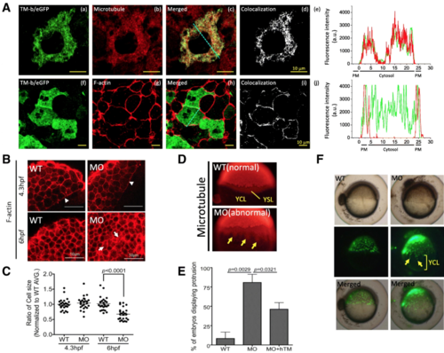
Cellular localization of zTM-b in developing embryos. a Wild-type embryos injected with plasmid zTM-b fused with eGFP (a, f; peGFP N1-zTM-b, 200 pg/embryo) at 1-cell stage were stained for microtubule (b) and F-actin (g) at 6 hpf with anti-α-tubulin antibodies and Alexa Flour 594 Phalloidin, respectively. (a-c, f-h) Confocal microscopy revealed distribution and colocalization of these proteins under excitation with a 488, or 543 nm laser. Blue dashed lines indicate the area of quantitative analyses of TM-b/microtubule/F-actin fluorescent intensity shown in (e) and (j). (d, i) Whole-cell pixel-by-pixel analyses of TM-b and microtubule or TM-b and F-actin colocalization. Scale bars, 10 μm. (e, j) Quantitative fluorescent analyses of cellular TM-b/microtubule/F-actin distribution. PM: plasma membrane. b, c Cell size was estimated from the dimension of blastomeres in the confocal images of phalloidin 594-stained embryos ( b). Actin signal was detected in cell peripheral and actin ring at the front edge of epibolic blastoderm (arrowheads). Increased signal dispersed in cytoplasm was also observed (arrows). For calculating cell dimension, total of 25 cells selected from 5 images for each group were analyzed with the on-line software Image J. Reported are the relative cell size normalized with the averaged size of wild-type blastomeres of the same stages. d, e Wild-type embryos injected with TM-b MO, with/without hTM rescue, were immuno-stained with anti-α-tubulin antibodies at 4.3 hpf. Embryos displaying signal with spike-like protrusion into YCL (arrows) were observed in zTM-b knockdown group and quantified. f Embryos at 4 hpf were injected with SYTOX green (0.25 mM/2.3 nL/embryo), a vital dye specific for nucleus staining, at the junction of blastoderm and yolk. The injected embryos were observed continuously under a dissecting fluorescence microscope and imaged at 6 hpf. Some nucleuses (arrow) within YSL moved ahead of the margin of blastoderm in TM-b morphants. Images were taken from lateral view with the animal pole to the top. YCL, yolk cytoplasmic layer; YSL, yolk syncytial layer; WT, wild-type embryos; MO, zTM-b morphants; hTM, human thrombomodulin |
| Fish: | |
|---|---|
| Knockdown Reagent: | |
| Observed In: | |
| Stage Range: | Dome to Shield |