
Fig. S4
- ID
- ZDB-FIG-190919-31
- Publication
- Shainer et al., 2019 - Agouti-Related Protein 2 Is a New Player in the Teleost Stress Response System
- Other Figures
- All Figure Page
- Back to All Figure Page
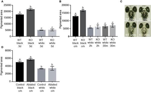
Aggregation and dispersion of melanosomes is normal in agrp2 KO and AgRP2 cell-ablated larvae. Related to Figure 4. (A) The pigmented area of 6-dpf larvae placed on a black or white background for 3 days. Pigmented area in agrp2 KO larvae was significantly higher on a black background, but did not differ when raised on a white background where both genotypes demonstrated equal aggregation of melanosomes (two-way ANOVA, n=25, p<0.01). Values represent mean pigmented area ± SE. (B) The pigmented area of 6-dpf larvae placed on a black background overnight (o/n) and then transferred to a white background for either 2 hours or 30 minutes, or kept on black background. On a black background, pigmented area was significantly higher in agrp2 KO, but when transferred to white background, both genotypes demonstrated equal aggregation of melanosomes, whether the adaptation was for 2 hours or 30 minutes (two-way ANOVA, n=30-35, p<0.01). Values represent mean pigmented area ± SE. (C) Representation of agrp2 KO and WT control melanosomes after 12 hours on black background, or after 30 minutes on white background. (D) The pigmented area of 6-dpf larvae placed on a black or white background overnight (o/n). Both AgRP2 ablated and their control larvae demonstrated equal aggregation or dispersion of melanosomes (two-way ANOVA, n=9, p<0.01). Values represent mean pigmented area ± SE |