
Fig. S2
- ID
- ZDB-FIG-190910-3
- Publication
- Diaz-Verdugo et al., 2019 - Mating Suppresses Alarm Response in Zebrafish
- Other Figures
- All Figure Page
- Back to All Figure Page
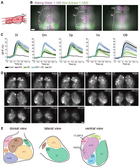
Widefield calcium imaging of olfactory bulb and telencephalon reveals brain area responding to mating water and skin extract. Olfactory bulb anatomy. Related to Figure 2. (A) Schematic representation of the forebrain of a juvenile zebrafish brain showing the anatomical position of the three optical sections imaged. (B) Mean ΔF/F signal in response to mating water (magenta) and skin extract (green; n = 5 fish). From left to right, in order from dorsal to ventral: three imaged optical sections. Solid magenta and green lines denote prominent areas that responded primarily to mating water and skin extract, respectively. (C) Mean ΔF/F signal time course in five representative areas (Dl, Dm, Dp, Vs and OB) for each odor delivered. Shaded regions represent the standard error of the mean. (D) Anatomical reference of 2-photon imaged olfactory bulb in 34 day-old juvenile tuba1a:GCaMP6s zebrafish. Number at the top right corner indicates the imaging depth. E) Schematic representation of the odor map and glomerular clusters of the olfactory bulb. Figure adapted from Yoshihara, 2014 [S1]. Abbreviations: Telencephalon (Tel), olfactory bulb (OB), lateral zone of the dorsal telencephalic area (Dl), medial zone of the dorsal telencephalic area (Dm), posterior part of the dorsal telencephalic area (Dp), and supracommisural nucleus of the ventral telencephalic area (Vs), dorsal glomerular (dG), dorso-lateral glomerular (glG), lateral (lG), medio-anterior (maG), medioposterior (mpG), medio-dorsal (mdG), ventro-anterior (vaG), ventro-medial (vmG), ventroposterior (vpG) glomerular cluster. Scale = 50 μm. |