
Fig. 1
- ID
- ZDB-FIG-190909-15
- Publication
- Bailey et al., 2019 - NAD+ improves neuromuscular development in a zebrafish model of FKRP-associated dystroglycanopathy
- Other Figures
- All Figure Page
- Back to All Figure Page
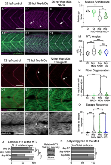
NAD+ or EmergenC supplementation at gastrulation improves muscle structure and function in fkrp morphants. (A–C) Anterior left, dorsal top, side-mounted embryos at 26 hpf stained for laminin-111 (purple) and actin (phalloidin, green). (A, A1) Control embryo. Laminin is concentrated at the MTJ. (B, B1) fkrp morphant. Although laminin is present at the MTJ (white arrow), it is also present within the myotome (white arrowhead). (C, C1) fkrp morphant treated with 0.1 mM NAD+ at 6 hpf. Laminin is concentrated at the MTJ (white arrow) as in control embryos. (D–F1) Anterior left, dorsal top, side-mounted embryos at 72 hpf stained for beta-DG (red) and f-actin (phalloidin, green). (D, D1) Control embryo. (E, E1) fkrp morphant. Beta-DG staining appears weaker at the MTJ (white arrowhead). (F, F1) fkrp morphant treated with EmergenC at 6 hpf. Beta-DG staining appears stronger at the MTJ (white arrow). (G–I) Anterior left, dorsal top, side-mounted embryos at 72 hpf stained for f-actin (phalloidin, gray). (G) Control embryo. (H) fkrp morphant. White arrowheads indicate single detached fibers. (I) fkrp morphant treated with EmergenC at 6 hpf. Note the improved muscle fiber structure. (J) Relative staining intensity of laminin-111 at the MTJ in 26 hpf embryos, based on no staining/localization (none, white), weak staining/localization (weak, light gray), and strong staining/localization (strong, dark gray) (images were blinded prior to analysis, see the “Methods” section). Controls (n = 23 embryos) have greater laminin intensity staining than fkrp morphants (n = 32 embryos). NAD+ (n = 10 embryos) and EmergenC (n = 30 embryos) supplementation improve laminin-111 concentration at the MTJ in fkrpmorphants. (K) Relative staining intensity of beta-DG at the MTJ in 72 hpf embryos, based on weak staining (weak, light gray) and strong staining (strong, dark gray). Although fkrp morphants have more embryos with weaker beta-DG staining at the MTJ (n = 7 embryos), there is no significant difference in beta-DG at the MTJ between untreated morphants, morphants treated with NAD+ (n = 7 embryos), EmergenC (n = 7 embryos), or control morphants (n = 5 embryos). (L) Quantification of muscle organization at 72 hpf. The anisotropy factor in embryos injected with fkrpMOs (n = 96 half-myotomes) is reduced compared to uninjected controls (n = 16 half-myotomes), and NAD+ (n = 40 half-myotomes) significantly increases the anisotropy factor in fkrp morphants. (M) Quantification of MTJ angles at 72 hpf. Injection of fkrp MOs (n = 343 MTJs) significantly increases MTJ angles compared to uninjected controls (n = 98 MTJs). Either NAD+ (n = 105 MTJs) or EmergenC (n = 158 MTJs) treatment significantly reduces MTJ angles compared to untreated morphants. (N) Untreated fkrp morphants (n = 54 embryos) have a significantly higher percent of myotomes per embryo with fiber detachments than controls (n = 40 embryos) at 72 hpf. NAD+ (n = 31 embryos) supplementation significantly reduces the percent of myotomes with dystrophy per embryo. (O) Injection of fkrpMOs (n = 184 embryos) significantly increases the number of touches required to induce an escape response compared to uninjected controls (n = 185 embryos). NAD+ (n = 84 embryos) or EmergenC (n = 111 embryos) treatment significantly reduces the number of touches to invoke an escape response. Scalebars are 50 μm. *p < 0.05, **p < 0.01, ***p < 0.001, ns non-significant |
| Fish: | |
|---|---|
| Conditions: | |
| Knockdown Reagent: | |
| Observed In: | |
| Stage: | Protruding-mouth |