
Fig. 4
- ID
- ZDB-FIG-190826-8
- Publication
- Kague et al., 2019 - Scleraxis genes are required for normal musculoskeletal development and for rib growth and mineralization in zebrafish
- Other Figures
- All Figure Page
- Back to All Figure Page
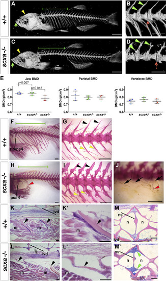
Adult scxa homozygous mutants have skeletal abnormalities. A–E) Three-dimensional volumetric isorenders from µCt data of scxa−/− and wild-type sibling showed absence of mineralized ribs (green dashed line above the rib region) (C, compared to A), and protruding jaws (green arrows) in mutants. Thoracic ribs region was magnified to show details of the vertebrae (B, D). Small bony structures were observed branching from the haemal arches (green arrowheads), and vertebrae misalignments were often present (red dashed arrow) in scxa mutants. E) BMD values were calculated from 3 distinctive bones: jaw (green arrows in A, C), vertebrae, and the parietal bone (yellow arrowhead in A, C). One-way ANOVA statistics with Tukey’s post hoc test performed, P values are as indicated. F–J) AR-stained adult scxa−/− mutants show no signal in ribs (H, under green line, compared with F), and neural (black arrowheads) and haemal arches (yellow arrowheads) have extensive bony growth (vertebrae 15–19 magnified in G, I). Fibrous, almost transparent ribs are seen in high magnification (black arrows, J), with the odd mineralized rib tissue (red arrowhead in H, J). The area rostrally to rib 5 is formed normally. Ps/r4, parapophysis and rib 4. K–M) Adult wild-type and scxa mutant zebrafish sagittal (KK′,LL′) and transversal (MM′) paraffin sections stained with H&E and AB. Existing rib structure in scxa mutant is short, thin, and wavy. The IVD is shown in MM′. The notochord string (ns) connecting the dorsal and ventral of the V shape is normal. However, the IVD is enriched with fibrous tissue (more purple, green arrowheads). M, muscle; n, notochordal cells. Scale bars, 1 mm (A–D, F–J), and 100 µm (K–M). |
| Fish: | |
|---|---|
| Observed In: | |
| Stage: | Adult |