
Fig. S10
- ID
- ZDB-FIG-190820-44
- Publication
- Zhu et al., 2019 - Aplnra/b Sequentially Regulate Organ Left-Right Patterning via Distinct Mechanisms
- Other Figures
- All Figure Page
- Back to All Figure Page
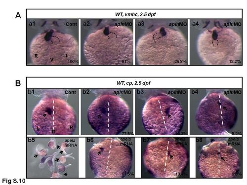
The organ LR patterning defect in apln morphants and apela mRNA injected embryos. (A. a1-a4) The heart LR patterning in control and apln MO injected embryos. In control embryos, 100% of embryos displayed normal heart loop (n=112). In apln morphants, 61%, 26.8% and 12.2% of embryos displayed normal, reversed and linear heart (A. a2-a4, n=41). For liver LR patterning, 97.3% of control embryos displayed left liver (B. b1, n=112). In apln morphants, 77.5%, 14.3% and 8.2% of embryos displayed left-sided, right-sided and middle/both sided liver (B. b2-b4, n=49). In embryos injected with apela mRNA, most of them displayed abnormal development (B. b5, arrow showed), 61.5%, 18.4% and 9.8% of embryos displayed left-sided, right-sided and middle/both sided liver (B. b6-b8, n=103). |