
Fig. S6
- ID
- ZDB-FIG-190812-41
- Publication
- Chen et al., 2019 - Cerebrovascular Injuries Induce Lymphatic Invasion into Brain Parenchyma to Guide Vascular Regeneration in Zebrafish
- Other Figures
- All Figure Page
- Back to All Figure Page
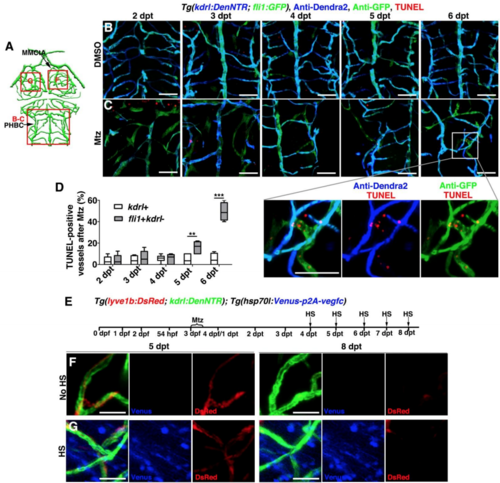
Apoptosis of the ingrown LECs after cerebrovascular regeneration, Related to Figures 6. (A) Illustrations of lower middle layer brain vascular network indicate the image areas (red boxes) in panels. (B–D) The TUNEL signals at 2-6 dpt after DMSO (B, n=16/18, 15/16, 16/19, 17/18, 15/16) or Mtz treatment (C, n=17/18, 14/17, 16/17, 15/18, 15/17). Note that the TUNEL signals gradually increased in the ingrown LECs (fli1+kdrl-) from 5 dpt onward. The statistics show the ratios of the TUNEL-positive vessels after Mtz treatment (D, n=6, Two-way ANOVA by Dunnett’s multiple comparisons test. kdrl+, 5 dpt vs fli1+kdrl-, 5 dpt, P=0.0025; kdrl+, 6 dpt vs fli1+kdrl-, 6 dpt, P<0.0001). Data are represented as mean ± SEM. ***P<0.001, **P<0.01. Ventral view, anterior upward. Scale bar, 50 μm. (E–G) Vegfc overexpression could not inhibit the clearance of ingrown lymphatic vessels. Transgenic lines, time points of Mtz treatment and heat-shock were shown in (E). Similar to the control larvae without heat-shock (F, n=9/9), overexpression of Vegfc by heat-shock was ineffective to clearance of lymphatic vessels at 8 dpt (G, n=10/12). Dorsal view, anterior upward. Scale bar, 20 μm. MMCtA, middle mesencephalic central artery; PHBC, primordial hindbrain channel. |
Reprinted from Developmental Cell, 49(5), Chen, J., He, J., Ni, R., Yang, Q., Zhang, Y., Luo, L., Cerebrovascular Injuries Induce Lymphatic Invasion into Brain Parenchyma to Guide Vascular Regeneration in Zebrafish, 697-710.e5, Copyright (2019) with permission from Elsevier. Full text @ Dev. Cell