
Fig. 9
- ID
- ZDB-FIG-190724-21
- Publication
- Seda et al., 2019 - An FDA-Approved Drug Screen for Compounds Influencing Craniofacial Skeletal Development and Craniosynostosis
- Other Figures
- All Figure Page
- Back to All Figure Page
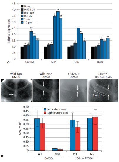
Functional assessment of calcineurin inhibition in skeletogenesis. A Relative expression of osteoblast differentiation marker genes normalised to untreated cells. * p < 0.05, **p < 0.005 (t test). B E18.5 calvarial explants cultured for 2 weeks in the presence or absence of 100 nMFK506. Top row: Low-power view of a control explant; note overlap of the midline parietal bones (arrows), showing continued osteogenesis of the sutures (1st and 2nd images). Whereas the DMSO-treated C342Y/+ coronal sutures undergo synostosis, FK506-treated C342Y/+ explants do not (arrows, 3rd and 4th images). The last image shows how the area of overlap of the frontal and parietal bones was measured over a fixed 1-mm length of suture (outlined) to gain a robust measure of suture width. These values are summarised over 33 explants, either wild-type (WT) or mutant (Mut), treated with DMSO or FK506. Values are given for both the left and right sutures for each explant. |