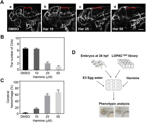
Inhibition of DYRK1A by harmine induces brain hemorrhage and vasculature defects in the hindbrain. (A) WT Tg(kdrl:EGFP) embryos were treated with increasing concentrations of DYRK1A inhibitor harmine, from 24 hpf until 52 hpf: Aa, DMSO; Ab, 10 µM harmine; Ac, 25 µM harmine; Ad, 50 µM harmine. Vascular patterning defects of CtAs are shown (red brackets) by confocal imaging at 52 hpf (lateral view). (B) Quantification of the effect on number of developing CtAs by harmine treatment. The numbers of CtAs were dramatically reduced by treating harmine in a dose-dependent manner. n=11 each group. (C) Quantification of the brain hemorrhage penetrance. The cerebral hemorrhagic phenotype of 2.3% in DMSO-treated embryos was increased from 14.8% to 65.7% by harmine treatment from 10 µM to 50 µM. The mean percentage for each treatment was shown from three independent experiments with approximately 40 embryos in each repeat. (D) Schematic showing the strategy of the in vivo chemical library screening to identify small molecule modifiers for cerebral hemorrhagic phenotype upon DYRK1A inhibition. Data are mean±s.e.m. Scale bar: 100 µm.
|
