|
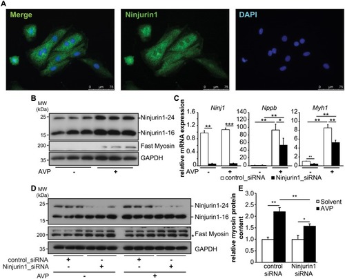
Knockdown of Ninjurin1 by siRNA leads to impaired cardiomyocyte hypertrophy <italic>in vitro</italic>.(A) Immunofluorescent staining of H9c2 myocytes with anti-Ninjurin1 as primary antibody and Alexa Fluor 488 conjugated secondary antibody (green). Nuclei were stained with DAPI (blue). Scale bar, 75 μm. (B) Hypertrophy of 7 days differentiated H9c2 myotubes was induced by arginine-vasopressin (AVP) treatment for 24 hours. Non-treated cells were used as control. Immunoblots of proteins isolated from H9c2 myotubes (as indicated) using anti-Ninjurin1 and anti-fast myosin antibody. GAPDH was used as loading control. The 16kDa (Ninjurin1-16) and 24kDa (Ninjurin1-24) Ninjurin1 isoforms are indicated. (C-E) 7 days differentiated H9c2 myotubes were transfected with control siRNA (control_siRNA) (n = 6) or siRNA targeting Ninjurin1 (Ninjurin1_siRNA) (n = 6). 48 hours after transfection hypertrophy was induced by AVP treatment for 24 hours (n = 3). Non-treated cells were used as control (n = 3). (C) Quantitative RT-PCR analysis of Ninjurin1 (Ninj1), Nppb and Myosin heavy chain 1 (Myh1). GAPDH expression was used as reference. Data are presented as mean ± SD. * P < 0.05, ** P < 0.01, *** P < 0.001(D) Immunoblots of proteins isolated from H9c2 myotubes (as indicated) using anti-Ninjurin1 and anti-fast myosin antibody. GAPDH was used as loading control. The 16kDa (Ninjurin1-16) and 24kDa (Ninjurin1-24) Ninjurin1 isoforms are indicated. (E) Densitometric analysis of fast myosin protein content from (D). Data are presented as mean ± SD. * P < 0.05, ** P < 0.01. MW, molecular weight; kDa, kilo Dalton. A two-tailed, unpaired Student’s t-test was used to calculate the P values.
|