
Figure 2
- ID
- ZDB-FIG-190723-2016
- Publication
- Mehta et al., 2014 - The Cellular and Physiological Functions of the Lowe Syndrome Protein OCRL1
- Other Figures
- All Figure Page
- Back to All Figure Page
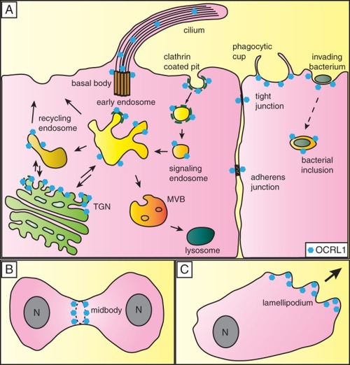
Cellular localization of OCRL1. A) OCRL1 (blue hexagons) has been localized to a number of cellular compartments. It is present at the TGN and various compartments of the endocytic pathway, where it resides at late-stage clathrin-coated pits, clathrin-coated vesicles, signaling endosomes, early or sorting endosomes and on recycling endosomes. OCRL1 has also been localized to the basal body of primary cilia, and it may also localize to the cilium itself. In maturing epithelia, OCRL1 transiently localizes to adherens and tight junctions. OCRL1 is recruited to phagosomes at a late stage in their formation, and is important for closure of the phagocytic cup as well as signaling events that occur post-sealing. OCRL1 is recruited to phagosomes generated by invading pathogenic bacteria such as |