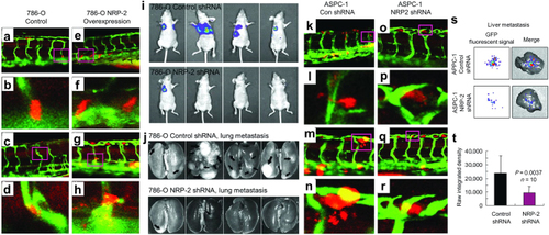
Human cancer cell xenograft models of extravasation in zebrafish and metastasis in mice. a–h Human 786-O renal cell carcinoma cells overexpressing retroviral control vector (a–d) or neuropilin-2 (NRP-2; e–h) were transiently labeled with cell tracker orange dye, microinjected into the pericardium of 3 dpf Tg(Fli-GFP) zebrafish, and imaged 1 day later. a–d control 786-O cells stay in the ISVs. e, f 786-O cells overexpressing NRP-2 extravasate from the ISVs. i–j 2 × 10⁶ luciferase-labeled 786-O cells suspended in PBS were subcutaneously injected into the right flank of female nude mice. Prior to the tumor growing to 10% of body weight, the subcutaneous tumors were surgically resected. Luciferase imaging was performed on the mice for 4–6 months to monitor metastasis, and the 786-O NRP-2 knockdown group (top) exhibited significantly fewer lung metastases than the control cohort (bottom). k–r Human ASPC-1 pancreatic cancer cells were transduced with control shRNA (k–n) or NRP-2 shRNA (o–r), transiently labeled, microinjected, and imaged as described above. k, l Extravasated control shRNA ASPC-1 cells. m, l Actively extravasating control shRNA ASPC-1 cells. o–r NRP-2 knockdown ASPC-1 cells stay in the ISV. s–t Male SCID mice were orthotopically injected with 2 × 10⁶ GFP-labeled ASPC-1 pancreatic cancer cells suspended in PBS, and after 15 days liver metastases were assessed by xenogen imaging. Reproduced with permission and adapted from: Cao Y. et al. Cancer Res 73, 4579–4590 (2013)84
|
