FIGURE

Figure 6

ID
ZDB-FIG-190723-1719
Publication
Ouyang et al., 2019 - CPSF1 mutations are associated with early-onset high myopia and involved in retinal ganglion cell axon projection
Other Figures
All Figure Page
Back to All Figure Page
Figure 6

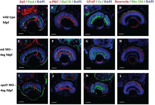
Retinal cells developed normally in WT larvae (A-D), std MO larvae (E-H) and cpsf1 morphants (I–L). (A, E, I) Co-labelling with anti-Pax6 (for ganglion and amacrine precursor cells, green) and anti-Zn5 (for mature RGCs and their axons, red) antibodies showed that the RGCs were mature. (B, F, J) Co-labelling with anti-HuC/D (for ganglion cells and amacrine cells, green) and anti-α PKC (for bipolar cells, red) antibodies indicated that the amacrine cells and bipolar cells developed normally. (C, G, K) Co-labelling with anti-GS (for Müller cells, green) and anti-GFAP (for glial cells and their axons, red) antibodies demonstrated that the Müller cells and glial cells had attained maturity. (D, H, L) Co-labelling with anti-Rho 1D4 (for long double-cone outer segments, green) and anti-Recoverin (for cone bipolar cells, red) antibodies demonstrated that the photoreceptors grew normally.

Expression Data
Antibodies:
Fish:
Knockdown Reagent:
Anatomical Terms:
Stage: Day 5

Expression Detail
Antibody Labeling
Phenotype Data

Phenotype Detail
Acknowledgments
This image is the copyrighted work of the attributed author or publisher, and ZFIN has permission only to display this image to its users. Additional permissions should be obtained from the applicable author or publisher of the image. Full text @ Hum. Mol. Genet.