
Figure 1
- ID
- ZDB-FIG-190723-1238
- Publication
- Starnes et al., 2012 - Neutrophil Reverse Migration Becomes Transparent with Zebrafish
- Other Figures
- All Figure Page
- Back to All Figure Page
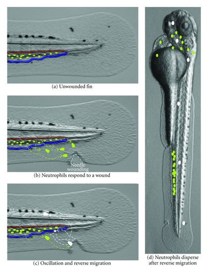
Neutrophil reverse migration in zebrafish larvae. This diagram illustrates the behavior of neutrophils undergoing reverse migration over an image of a 3-day postfertilization zebrafish larva that was PTU-treated to prevent pigmentation. (a) In unwounded larva, neutrophils (green ovals) are present in the caudal hematopoietic tissue (CHT), which is situated between the caudal artery (red shading) and the caudal vein (blue shading). (b) In response to a wound, neutrophils are mobilized from the CHT, and they migrate through the tissue towards a wound. (c) The green to white color change represents the ability to photoconvert individual neutrophils that reach a wound. Neutrophils often migrate between the wound and the vasculature multiple times before finally departing. Neutrophils have been observed performing reverse migration by entering the vasculature and by migrating through tissues in zebrafish larvae. (d) Reverse migrated neutrophils (white) are found dispersed throughout the body without an obvious tissue preference 2 days after leaving wounds. |