
Fig. 4
- ID
- ZDB-FIG-190626-4
- Publication
- Verweij et al., 2019 - Live Tracking of Inter-organ Communication by Endogenous Exosomes In Vivo
- Other Figures
- All Figure Page
- Back to All Figure Page
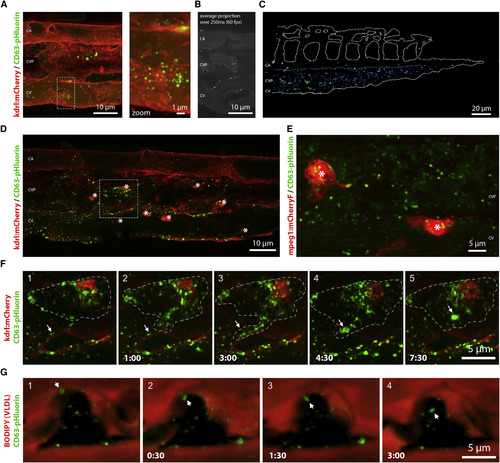
Distribution and Target Cells of YSL Exosomes (A) Still of time-lapse shown in Video S3A of the CVP of a 3 dpf Tg(kdrl:Hsa.HRAS-mCherry) embryo expressing CD63-pHluorin in the YSL. Dashed box indicates area shown at higher magnification to the right. (B) Average projection of 15 sequential frames of Video S3B, showing caudal artery (CA), CVP, and the cardinal vein (CV) in 3 dpf zebrafish embryos expressing CD63-pHluorin in the YSL. (C) Heatmap of CD63-pHluorin signal in CVP of 6 3 dpf Tg(kdrl:Hsa.HRAS-mCherry) zebrafish embryos expressing CD63-pHluorin in the YSL. (D) CVP of a 3 dpf Tg(kdrl:Hsa.HRAS-mCherry) embryo expressing CD63-pHluorin in the YSL. Stars indicate macrophages. Dashed box indicates area shown in Video S4B and (F). Asterisks indicate macrophages. (E) Still of Video S4A, showing the CVP of a 3 dpf Tg(mpeg1:mCherryF) expressing CD63-pHluorin in the YSL. Asterisks indicate macrophages. (F) Time-lapse of a macrophage in a 3 dpf Tg(kdrl:Hsa.HRAS-mCherry) zebrafish embryo expressing CD63-pHluorin in the YSL, shown in Video S4B. (G) Time-lapse of macrophage in a BODIPY-C12 injected 3 dpf zebrafish embryo expressing CD63-pHluorin in the YSL, shown in Video S4C. CA, caudal artery; YSL, yolk syncytial layer; CVP, caudal vein plexus. |
Reprinted from Developmental Cell, 48(4), Verweij, F.J., Revenu, C., Arras, G., Dingli, F., Loew, D., Pegtel, M.D., Follain, G., Allio, G., Goetz, J.G., Zimmermann, P., Herbomel, P., Del Bene, F., Raposo, G., van Niel, G., Live Tracking of Inter-organ Communication by Endogenous Exosomes In Vivo, 573-589.e4, Copyright (2019) with permission from Elsevier. Full text @ Dev. Cell