
Fig. 5
- ID
- ZDB-FIG-190625-5
- Publication
- Pacentine et al., 2019 - Subunits of the mechano-electrical transduction channel, Tmc1/2b, require Tmie to localize in zebrafish sensory hair cells
- Other Figures
- All Figure Page
- Back to All Figure Page
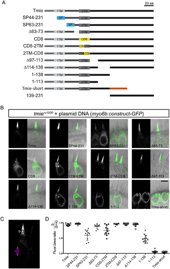
Schema for a systematic domain analysis of Tmie and subcellular localization of Tmie constructs. (A) A linear diagram of 13 unique constructs of Tmie used in our experiments. Full-length zebrafish Tmie contains two hydrophobic regions predicted to form transmembrane helices (1TM and 2TM). SP44-231 and SP63-231 replace part of the N-terminus with a signal peptide (SP) from the Glutamate receptor 2a (in blue). In the CD8, CD8-2TM, and 2TM-CD8 constructs, all or part of the 2TM is replaced by the helix from the CD8 glycoprotein (in yellow). Tmie-short is a fish-specific isoform of Tmie that contains an alternate final exon (in orange). Dotted lines represent internal deletions. (B) Representative confocal images of each construct being expressed as a GFP-tagged transgene in hair cells of 4–6 dpf tmieru1000 larvae. Expression is mosaic due to random genomic insertion into subsets of progenitor cells after single-cell injection. All images are of cells in the inner ear cristae. Scale bar is 5μm. (C) The localization of each GFP fusion protein was determined by measuring the fluorescence/area in the bundle (b) and soma (s), and then calculating b / (b+s). (D) Enrichment in the hair bundle is displayed as a ratio for each construct, with 1 being completely bundle-enriched and 0 being completely soma-enriched. |