
Fig. 6
- ID
- ZDB-FIG-190618-38
- Publication
- Hanovice et al., 2019 - Regeneration of the zebrafish retinal pigment epithelium after widespread genetic ablation
- Other Figures
- All Figure Page
- Back to All Figure Page
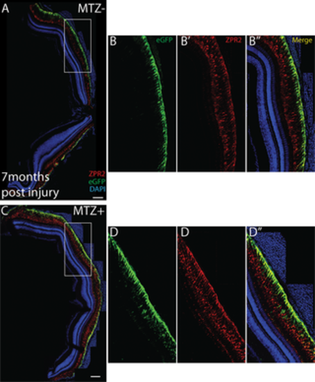
Regenerated RPE appears normal 7 months post-injury. Stitched-together confocal images of transverse cryosections of 7 month post-injury fish (C,D) and age-matched sibling controls (A-B). (A) Robust eGFP and ZPR2 expression exists throughout the central RPE. (B-B”) Within the central RPE, eGFP is strongly expressed in the RPE cell body and labels apical processes, most strongly toward the cell body, while ZPR2 labels the RPE cell body and apical processes closer to the outer limiting membrane. (C) In ablated fish, strong expression of eGFP and ZPR2 is evident throughout the retina. (D-D”), and a similar pattern of eGFP and ZPR2 expression is observed in the central injury site. Scale bar = 100μm. |
| Gene: | |
|---|---|
| Antibody: | |
| Fish: | |
| Condition: | |
| Anatomical Term: | |
| Stage Range: | Day 6 to Adult |
| Fish: | |
|---|---|
| Condition: | |
| Observed In: | |
| Stage Range: | Day 6 to Adult |