
Fig. 1
- ID
- ZDB-FIG-190523-2
- Publication
- Lalonde et al., 2018 - Contributions of 5'HoxA/D regulation to actinodin evolution and the fin-to-limb transition
- Other Figures
- All Figure Page
- Back to All Figure Page
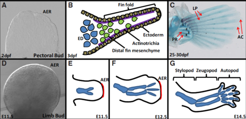
Overview of zebrafish pectoral fin and mouse limb skeletal development. (A-C) Zebrafish pectoral fin development at 2 days post fertilization (dpf), 3dpf and 25-30dpf. (D-G) Mouse forelimb development at E11.5, E12.5 and E14.5. At 2dpf, the zebrafish pectoral fin consists of a bud possessing an apical ectodermal ridge (A). At 3dpf, the fin fold is supported by actinotrichia (purple lines) and distal fin mesenchyme migrates though the fin fold using the actinotrichia as a scaffold (green cells) (B). The proximal mesenchyme condenses and chondrifies to form the endoskeletal disc (blue cells) (B). At 25-30dpf, the proximal radials are still composed of cartilage (numbered 1-4), and the lepidotrichia have started calcifying. Actinotrichia are restricted to the distal tip of each fin ray (C). The mouse forelimb starts with the formation of a bud very similar to the pectoral fin bud (D). From E11.5 to E.14.5 cartilaginous templates will form for the three limb segments: stylopod, zeugopod and autopod (E-G). The AER regresses after E12.5 in the mouse forelimb (F,G). AC, actinotrichia; AER, apical ectodermal ridge; ED, endoskeletal disc; LP, lepidotrichia; PR, proximal radials.
|