|
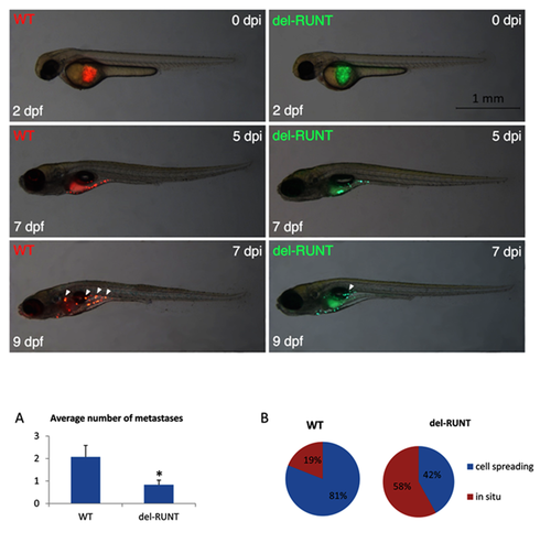
Differential migratory ability of wild-type and del-RUNT cells in zebrafish xeno-transplants. Representative images show wild-type (WT, stained in red) and del-RUNT (del-RUNT, stained in green) cells xeno-transplanted in zebrafish embryos 2 days post fertilization (dpf). At day 0 post injection (dpi), transplanted cells are still located in the yolk. At 5 dpi, cells appear more dispersed in the yolk of 7 dpf larvae. At 7 dpi/9 dpf, initial metastases (indicated by white arrowheads) are detectable in WT and, to a lesser extent, in del-RUNT- transplanted larvae. All views are lateral, anterior to the left. Charts on average number of metastases and cell spreading, calculated at 9 dpf in WT (N:22) and del-Runt (N:12) transplanted zebrafish, are shown in (A) and (B), respectively. * p < 0.05.
|