
Fig. 2
- ID
- ZDB-FIG-190201-25
- Publication
- Shaw et al., 2018 - Stable transgenic C9orf72 zebrafish model key aspects of the ALS/FTD phenotype and reveal novel pathological features
- Other Figures
- All Figure Page
- Back to All Figure Page
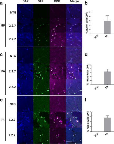
C9orf72 model zebrafish display DPR expression in the nucleus. (a,b) Immunofluorescence labelling of adult zebrafish muscle tissue showed that poly-GP DPR protein localises to the nucleus in 2.2–2 and 2.2–7 transgenic zebrafish. (c.d) Immunofluorescence labelling of adult zebrafish muscle tissue showed that poly-PA DPR protein localises to the nucleus in 2.2–2 and 2.2–7 transgenic zebrafish. (e,f) Immunofluorescence labelling of adult zebrafish muscle tissue showed that poly-PA DPR protein localises to the nucleus in 2.2–2 and 2.2–7 transgenic zebrafish. For all DPR images nuclei are stained with Hoechst (blue), GFP is stained with GFP antibody (green) and DPR proteins are stained with the relevant DPR antibody (purple), white arrow heads denote DPR positive staining. Scale bar = 25 μm for all DPR images |
| Gene: | |
|---|---|
| Fish: | |
| Anatomical Term: | |
| Stage: | Adult |