
Fig. 4
- ID
- ZDB-FIG-190110-1
- Publication
- Hunter et al., 2018 - Oxidative Stress Orchestrates Cell Polarity to Promote Embryonic Wound Healing
- Other Figures
- All Figure Page
- Back to All Figure Page
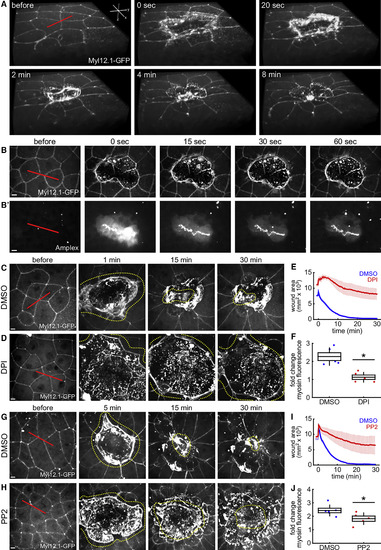
Wound Repair in Zebrafish Embryos Is ROS- and Src-Dependent (A) Volume rendering of wound closure in the enveloping layer (EVL) of a zebrafish embryo expressing non-muscle myosin light-chain 12-GFP (Myl12.1-GFP). (B and B′) Wound healing in the EVL of a zebrafish embryo expressing Myl12.1-GFP (B) and treated with the ROS dye Amplex UltraRed (B′). (C, D, G, and H) Wound closure in the EVL of a zebrafish embryo expressing Myl12.1-GFP and treated with 1% DMSO (C and G), and 250 μM DPI in 1% DMSO (D) or 20 μM PP2 in 1% DMSO (H). Yellow dotted lines outline wounds. (B–D, G, and H) Scale bars, 10 μm. (A–D, G, and H) Time after wounding is shown. Red lines indicate wound sites. (E, F, I, and J) Mean wound area over time (E and I) or mean myosin fold change in the visible segments of the wound margin -quantified 3 min after wounding to avoid the accumulation of apically extruded cellular debris- (F and J) for zebrafish embryos treated with DMSO (blue; E and F, n = 5; I and J, n = 6), DPI (red; E and F, n = 5), or PP2 (red; I and J, n = 5). Wound areas for DPI are underestimated due to wound overexpansion out of the field of view. (E and I) Error bars, SEM. (F and J) Horizontal line, mean; box, SEM; error bars, SD. ∗, p < 0.05. |
Reprinted from Developmental Cell, 47, Hunter, M.V., Willoughby, P.M., Bruce, A.E.E., Fernandez-Gonzalez, R., Oxidative Stress Orchestrates Cell Polarity to Promote Embryonic Wound Healing, 377-387.e4, Copyright (2018) with permission from Elsevier. Full text @ Dev. Cell