
Fig. S9
- ID
- ZDB-FIG-181207-36
- Publication
- Tessadori et al., 2018 - Effective CRISPR/Cas9-based nucleotide editing in zebrafish to model human genetic cardiovascular disorders
- Other Figures
- All Figure Page
- Back to All Figure Page
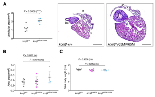
Adult kcnj8[V65M] fish show enlarged ventricular area. (A) Ventricular area in homozygous kcnj8[V65M] mutants. Representative heart histology of adult kcnj8V65M/V65M mutants and wildtype siblings after H&E staining. Exemplary depiction of 1 WT and 1 kcnj8[V65M] heart. For assessment of ventricular chamber size, tissue sections showing the largest ventricular area were selected and area was quantified using ImageJ (NIH). (B) Atrial area in heterozygous and homozygous kcnj8[V65M] mutants. For assessment of atrial chamber size, tissue sections showing the largest atrial area were selected and area was quantified using ImageJ (NIH). (C) Total body length in kcnj8[V65M] mutants. To account for variations in heart size overall body length was measured from the tip of the head to the end of the trunk (before the caudal fin). kcnj8+/+ controls are the same as in Fig.2E. For all graphs, significance was determined by twotailed unpaired Student's t test or Mann–Whitney two-tailed U test: * p≤0.05; ** p≤0.01; *** p≤0.001; **** p≤0.0001. The black horizontal bar indicates the mean value for each condition. Sample size, kcnj8+/+, n=6; kcnj8+/V65M, n=6; kcnj8V65M/V65M, n=6 in A-C. Scale bars, 500 μm in A. All embryos analyzed originated from group matings of adult zebrafish. |
| Fish: | |
|---|---|
| Observed In: | |
| Stage: | Adult |