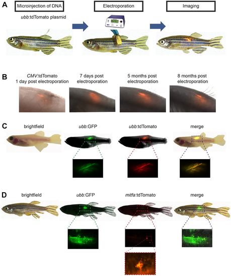
Fig. 1
- ID
- ZDB-FIG-181109-31
- Publication
- Callahan et al., 2018 - Cancer modeling by Transgene Electroporation in Adult Zebrafish (TEAZ)
- Other Figures
- All Figure Page
- Back to All Figure Page
|
TEAZ. (A) Schematic representation of the method applied for the introduction of ubb:tdTomato directly under the dorsal fin of adult zebrafish. The purified plasmid DNA (1 μl of a 1000 ng/μl solution of ubb:tdTomato) is injected into anesthetized zebrafish using a pulled glass micropipette. Electrical pulses are directed across the injected region (settings: LV mode, 45 V, 5 pulses, 60 ms pulse length and 1 s pulse interval). Reporter expression can be visualized by fluorescent microscopy (n=2/2). (B) Electroporation of a CMV:tdTomato plasmid was performed and the animal followed for a period of 8 months (n=2/2). The fluorescent signal can be visualized as early as 1 dpe, with intensity peaking at ∼1 week and maintaining for at least 8 months. (C) Multiple plasmids will co-integrate in TEAZ. casper zebrafish were electroporated with a total volume of 1.0 μl (0.5 μl of 1000 ng/μl ubb:GFP and 0.5 μl of 1000 ng/μl ubb:tdTomato) and imaged using BF, GFP and tdTomato (n=3/3), revealing co-expression of the plasmids. (D) Promoter specificity is maintained following TEAZ. AB fish were electroporated with 1.0 μl total volume (0.5 μl of 1000 ng/μl ubb:GFP and 0.5 μl of 1000 ng/μl mitfa:tdTomato) and displayed highly restricted expression of the mitfa reporter plasmid, but widespread expression of the ubb plasmid (n=9/9). High-resolution imaging of the tdTomato+ cells reveals a dendritic phenotype, consistent with the melanocytic lineage. |

