
Fig. S6
- ID
- ZDB-FIG-180913-63
- Publication
- Asakawa et al., 2018 - Protocadherin-Mediated Cell Repulsion Controls the Central Topography and Efferent Projections of the Abducens Nucleus
- Other Figures
- All Figure Page
- Back to All Figure Page
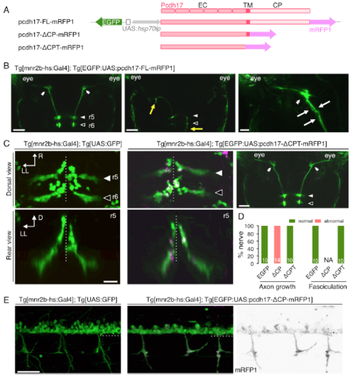
Ectopic expression of the truncated Pcdh17 proteins in the abducens and spinal motor neurons. (Related to Figure 6) (A) UAS constructs for expression of the full-length and truncated Pcdh17 proteins. (B) Normal (left), misguided (middle, yellow arrows) and defasciculated (right, white arrows) axon projections of mnr2b-ABNs expressing Pcdh17-FL-mRFP1 at 3 dpf. The bars indicate 50 μm in left, middle and 20 μm in right. (C, D) pcdh17-ΔCPT expression does not affect soma topography and axon projection of mnr2b-ABNs at 3 dpf. Numbers of the nerves investigated are shown in the bar (D). The bars indicate 20 μm (left) and 50 μm (right). R, rostral; LL, left lateral, D, dorsal. (E) pcdh17-ΔCP expression causes clumping of somas and axon projection of spinal motor neurons at 3 dpf. The dashed lines indicate the ventral limit of the spinal cord. The bar indicates 100 μm. |