
Fig. 3
- ID
- ZDB-FIG-180912-51
- Publication
- Jin et al., 2018 - Foxi1 promotes late-stage pharyngeal pouch morphogenesis through ectodermal Wnt4a activation
- Other Figures
- All Figure Page
- Back to All Figure Page
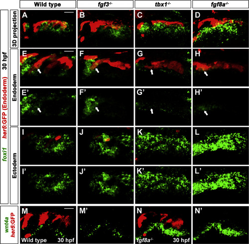
Requirements for Tbx1 and Fgf8a in repression of the ectodermal Foxi1 and Wnt4a. (A-L) Fluorescent in situ hybridization for foxi1 (green) and GFP immunohistochemistry to detect her5:GFP-positive endoderm (red) at 30 hpf. (A, B) In wild-type embryos and fgf3 mutants, foxi1 expression was observed in her5:GFP-positive endoderm (arrows in E and F) as well as in facial ectoderm (I and J). (C) In tbx1 mutants, foxi1 expression was significantly decreased in the her5:GFP-positive endoderm (arrow in G), whereas it was expanded ectopically in the facial ectoderm (K). (D) In fgf8a mutants, foxi1 expression was detected in the her5:GFP-positive endoderm (arrow in H) and foxi1 expression in the facial ectoderm was expanded ectopically and increased in intensity (also see L). (E-G) Higher magnification image of wild- type embryos and mutants focusing on the her5:GFP-positive endoderm. (I-L) Higher magnification image of wild-type embryos and mutants focusing on the facial ectoderm. (M, N) Fluorescent in situ hybridization for wnt4a (green) and GFP immunohistochemistry to visualize her5:GFP-positive pouches (red) at 30 hpf. wnt4a expression was observed in ectodermal patches in wild-type embryos (M), whereas it became stronger and was expanded ectopically in the facial ectoderm of fgf8a mutants (N). (E′-N′) Green channel only. Scale bars: 40 µm (A-D, M, and N), 20 µm (E-L). |
Reprinted from Developmental Biology, 441(1), Jin, S., Jiyun, O., Stellabotte, F., Choe, C.P., Foxi1 promotes late-stage pharyngeal pouch morphogenesis through ectodermal Wnt4a activation, 12-18, Copyright (2018) with permission from Elsevier. Full text @ Dev. Biol.