
Fig. S13
- ID
- ZDB-FIG-180906-26
- Publication
- Mochizuki et al., 2017 - Cell division and cadherin-mediated adhesion regulate lens epithelial cell movement in zebrafish
- Other Figures
- All Figure Page
- Back to All Figure Page
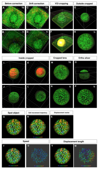
Procedure for obtaining 3D time-lapse images of anterior lens epithelium (A–D) Two time points from T0 to Ti of embryonic eyes without (A, B) and with drift correction (C, D). (E, F) VOI cropping by drawing outside contour circles and creation of a surface masking the lens. (G, H) Outside cropped lens. (I–L) Cropping of the lens fiber area by drawing inside contour circles and by creating a surface masking the lens fiber area. (M, N) Cropped lens at T0 and Ti. (O, P) Extract of anterior lens epithelium using the “Ortho Slicer” tool. (Q–W) Analysis of cell movement of lens epithelial cells. (Q) Assignment of spot objects to trace cell lineages, which classify epithelial cells into dividing (yellow), non-dividing (blue), and eliminated cell populations (purple). (R) Cell movement trajectory, (S) displacement vector, (T–U) cell movement speed, and (V–W) displacement length. |