
FIGURE
Fig. 4
- ID
- ZDB-FIG-180716-17
- Publication
- Ishii et al., 2018 - Genetic Requirement of talin1 for Proliferation of Cranial Neural Crest Cells during Palate Development.
- Other Figures
- All Figure Page
- Back to All Figure Page
Fig. 4
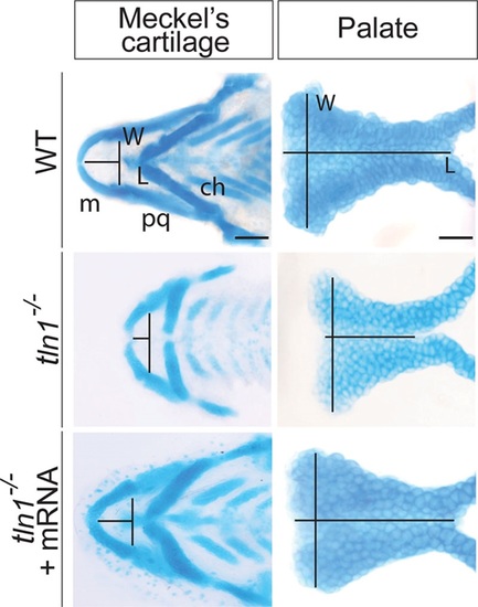
Alcian blue images of flat mounted Meckel’s cartilage and the palate of the WT (above), tln1 (center), and rescued mutants (below) at 4 dpf are shown. The length (L) of the Meckel’s cartilage is measured from the midline of the Meckel’s cartilage to the midline of an imaginary line drawn joining the joints between the Meckel’s cartilage and the palatoquadrate. Likewise the width (W) is across the joints between the Meckel’s cartilage and the palatoquadrate. The L of the palate is measured from the anterior to the posterior of the palate through its mid point, whereas W is measured as the maximum distance between the 2 lateral edges at the anterior most region. Tln1 mutants have a shorter Meckel’s cartilage (center and left) and palate (center and right). Injection of full-length tln1 mRNA partially rescues the phenotypes (below). Scale bars: 500 μm (Meckel’s cartilage) and 100 μm (palate). ch, ceratohyal; L, length; m, Meckel’s cartilage; pq, palatoquadrate; W, width.
|
Expression Data
Expression Detail
Antibody Labeling
Phenotype Data
| Fish: | |
|---|---|
| Observed In: | |
| Stage: | Day 4 |
Phenotype Detail
Acknowledgments
This image is the copyrighted work of the attributed author or publisher, and
ZFIN has permission only to display this image to its users.
Additional permissions should be obtained from the applicable author or publisher of the image.
Full text @ Plast Reconstr Surg Glob Open