
Fig. 5
- ID
- ZDB-FIG-180712-53
- Publication
- Lu et al., 2018 - 50 Hz volumetric functional imaging with continuously adjustable depth of focus
- Other Figures
- All Figure Page
- Back to All Figure Page
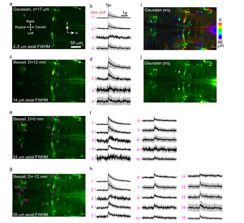
50 Hz volumetric functional calcium imaging of volumes of spinal projection neurons in zebrafish larvae. (a) Image acquired by Gaussian focus scanning at 127 μm from the dorsal surface of the head (relative depth z = 17 μm). (b) Averaged calcium transients of neurons evoked by the acoustomechanical tapping stimuli. (c), (e), and (g) were volumetric images obtained by scanning a short (14 μm axial FWHM), medium (24 μm axial FWHM), and long (39 μm axial FWHM) Bessel foci, respectively. (d), (f), and (h) were averaged calcium transients of responsive neurons. An acoustomechanical stimulus was delivered at 0 s. The Gaussian focus had 2.6 μm axial FWHM. The short (displacement of lens L2 D = 12 mm), medium (D = 0 mm) and long Bessel (D = −12 mm) foci has 14 μm, 24 μm and 39 μm axial FWHMs. The field of view was 366 μm × 214 μm. (See Visualization 2 for the functional movies.) (i) and (j) Mean intensity projections of a 66-μm-thick image stack acquired by Gaussian focus scanning (see Visualization 3 for the Gaussian 3D stack). Color in (i) encodes relative depth. Eleven trials were averaged in (b), (d), (f), and (h). Shadow represents standard deviations. Post-objective power: (a) 38 mW, (c) 97 mW, (e) 110 mW and (g) 132 mW. |