
Fig. 4
- ID
- ZDB-FIG-180712-43
- Publication
- Shibata et al., 2018 - Heterogeneous fates and dynamic rearrangement of regenerative epidermis-derived cells during zebrafish fin regeneration
- Other Figures
- All Figure Page
- Back to All Figure Page
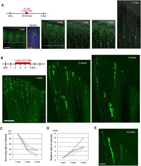
Later emerging RE cells contribute to the regenerated epidermis. (A) Tracking of the progeny of fn1b-expressed RE cells that were labelled by Cre-loxP recombination at 23-24 hpa in the Tg(fn1b:creERt2;Olactb:loxP-dsred2-loxP-egfp). A longitudinal section of the 2 dpa fin shows that EGFP+ cells are found in both basal and non-basal cells in the RE. The section was counterstained with DAPI. The dotted line indicates the amputation plane. Scale bars: 200 µm (fins) and 50 µm (section). (B) Tracking of the progeny of RE cells that were labelled by Cre-loxP recombination at 2-5 dpa. Cell colonies derived from the Cre-labelled cells were evident at 1 wpa; however, whereas the size of the colonies grew over time, the number of colonies decreased. (C) Quantification of the number of colonies derived from the Cre-labelled cells in B at 1, 2 and 3 wpa (n=4 zebrafish). Approximately 80% of the colonies disappeared between weeks 1 and 3. Each line in the graph corresponds to an individual fish. (D) Quantification of the size of colonies derived from the Cre-labelled cells in B at 1, 2 and 3 wpa (n=4 zebrafish). In contrast to the number of colonies, the colony size increased between 1 and 3 weeks. Each line in the graph corresponds to an individual fish. (E) Tracking of the Cre-labelled RE-derived cells after 4 months. Progeny of the RE-derived cells were still retained in the epidermis, although their number was decreased relative to the number at 3 wpa. Scale bar: 500 µm. |