
FIGURE
Fig. 4
- ID
- ZDB-FIG-180524-11
- Publication
- Saera-Vila et al., 2018 - Extraocular muscle regeneration in zebrafish requires late signals from Insulin-like growth factors
- Other Figures
- All Figure Page
- Back to All Figure Page
Fig. 4
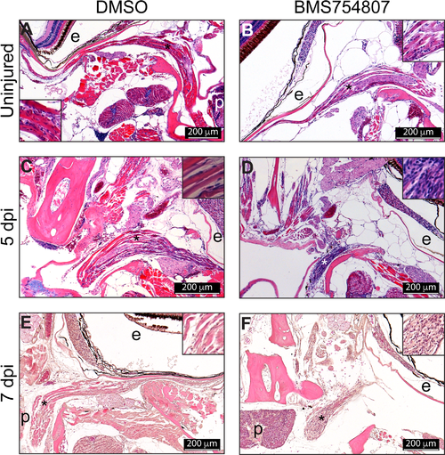
Histological analysis of the regenerating muscle. Paraffin sections (5 μm) H&E staining of regenerating muscles from DMSO (A, C) and BMS754807 (B, D) treated fish. Sections of regenerating muscle at 5 (A, B) and 7 dpi (C, D). The asterisk marks the approximate position of the inset. Images are representative examples from 5 fish analyzed per treatment and time point. P, pituitary; e, eye. The S1 Fig shows a zebrafish coronal section diagram as a reference for the position of the pictures shown in this figure. |
Expression Data
Expression Detail
Antibody Labeling
Phenotype Data
Phenotype Detail
Acknowledgments
This image is the copyrighted work of the attributed author or publisher, and
ZFIN has permission only to display this image to its users.
Additional permissions should be obtained from the applicable author or publisher of the image.
Full text @ PLoS One