
Fig. 3
- ID
- ZDB-FIG-180511-55
- Publication
- Weijts et al., 2017 - Atypical E2Fs inhibit tumor angiogenesis
- Other Figures
- All Figure Page
- Back to All Figure Page
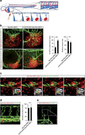
Absence of E2f7/8 in zebrafish xenograft tumors leads to increased tumor vascularization. (a) Schematic representation of xenograft positions and experimental setup. Labeled MEFs were injected 48 h post fertilization (hpf) in the perivitelline space on the yolk sac, ventrally of the sub intestinal vein (SIV) and posterior of the common cardinal vein (CCV or duct of Cuvier). Grafts tend to evoke an angiogenic response from the SIV, but frequently also from the CCV (3a’). In addition, neoplastic cells from the injection site are able to enter the circulation via the vessels formed by tumor angiogenesis and often attach or simply get stuck in the caudal vein (CV) region (3a’’). Reprinted with permission from Magliozzi, R. et al.31 (b) Representative images and quantification of tumor angiogenesis and size of control (Red; DiI; n=10) and E2f7/8 DKO tumors (Red; DiI; n=10) injected in Tg(fli1a:gfp) zebrafish. (c) Time-lapse series that shows metastasizing cell (dashed line) from the tumor into the vasculature. Insets display metastasizing cell only. (d) Representative image and quantification of MEFs metastasis in the trunk region. (e) Example of extravasation of (E2F7/8 DKO) MEFs into the surrounding tissue. Abbreviations: CV, caudal vein; CCV, common cardinal vein; hpi, hours post injection. All quantified data present the average±s.d. compared with the indicated controls in at least three independent experiments. ***P<0.05; ns, not significant. |
| Gene: | |
|---|---|
| Fish: | |
| Condition: | |
| Anatomical Terms: | |
| Stage: | Day 4 |