Fig. S3
- ID
- ZDB-FIG-180501-35
- Publication
- Banerji et al., 2017 - Cohesin mediates Esco2-dependent transcriptional regulation in zebrafish regenerating fin model of Roberts syndrome
- Other Figures
- All Figure Page
- Back to All Figure Page
|
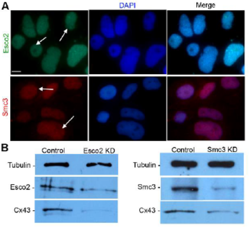
AB9 cells as a system to evaluate cohesin-binding at the cx43 promoter. (A) Expression of Esco2 and Smc3 are detected in AB9 cells by immunofluorescence. The anti-Esco2 antibody and anti-Smc3 antibody stains the nuclei of the cells (DAPI, blue), indicated with arrows. For each protein, 3 independent trials were performed. Scale bar is 10 μm (B) Western blot analysis detects the Esco2 in Std-MO electroporated control cell lysates (Lane1: Control) at a predicted size of 68 kDa. A reduction in Esco2 protein levels in MO1-electroporated esco2 knockdown cell lysate (lane 2: Esco2 KD) was observed when compared to the control sample (lane 1: Control). The results with anti-Cx43 antibody reveals reduced Cx43 protein levels (detected at 43 kDa as predicted) in Esco2 KD compared to control. Western blot analysis detects Smc3 in Std-MO electroporated control cell lysates (lane1: Control) at a predicted size of 142 kDa. A reduction in Smc3 protein levels in MO1-electroporated smc3 knockdown cell lysate (lane 2: Smc3 KD) was observed when compared to the control sample (lane 1: Control). The resultswith anti-Cx43 antibody reveals reduced Cx43 protein levels (detected at 43 kDa as predicted) in Smc3 KD compared to control. Tubulin detected at 50 kDa was used as the loading control for both blots. Image J software was used for analysis of relative band intensity from data of 3 independent trials |

