
Fig. 2
- ID
- ZDB-FIG-180412-15
- Publication
- Ribeiro et al., 2017 - Foxj1a is expressed in ependymal precursors, controls central canal position and is activated in new ependymal cells during regeneration in zebrafish
- Other Figures
- All Figure Page
- Back to All Figure Page
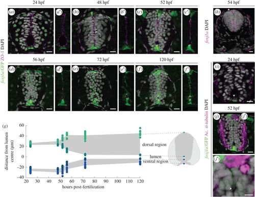
Foxj1a+ cells participate in the formation of the spinal cord central canal. (a–f′) Representative images of the neural tube region in transverse sections of Tg(0.6foxj1a:GFP) transgenic zebrafish embryos/larvae ranging from 24 to 120 hpf. The apical edge of the cells surrounding the lumen is identified by ZO-1 immunostaining (magenta) and the GFP reporter labels Foxj1a-expressing cells (green). (g) Quantification of lumen closure from 24 to 120 hpf. The positions of the floor plate, ventral and dorsal points of the lumen and roof plate are normalized to the middle point of the lumen. Sample number: 24 hpf (n = 12); 48 hpf (n = 6); 52 hpf (n = 9); 56 hpf (n = 9); 72 hpf (n = 11); 120 hpf (n = 8). (h) Confocal image of a FISH of foxj1a (magenta) in a 54 hpf embryo. foxj1a is expressed in the floor plate, ventro-lateral cells, roof plate (arrowhead) and pronephric (asterisks). (i,j,j′) Long cilia (arrowhead), labelled by acetylated α-tubulin (magenta), are present in Foxj1a-positive floor plate cells at 24 hpf (i) and in Foxj1a-expressing roof plate cells in the roof plate at 52 hpf (j,j′). DAPI-labelled nuclei are shown in grey. Scale bars: 10 µm in a–j; 5 µm in j′. |