
Fig. s2
- ID
- ZDB-FIG-180410-10
- Publication
- Nagashima et al., 2017 - Anisotropic Müller glial scaffolding supports a multiplex lattice mosaic of photoreceptors in zebrafish retina
- Other Figures
- All Figure Page
- Back to All Figure Page
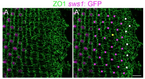
Immature UV cones have rounded apical profiles in the pre-column area. (A, A’) Retinal flat-mount immunocytochemistry for ZO1 (green) in a juvenile transgenic zebrafish with the UV cone reporter, Tg(sws1: EGFP) (magenta). The onset of sws1 opsin transgene expression is delayed in the differentiating UV cones until after the organization of cone columns. (A’) The precision of the mosaic pattern allows profiles that are Tg(sws1: EGFP)-negative to be identified as UV cones (magenta dots), and the position and shape of the large rounded profiles in the pre-column zone (white dots) suggests that these, too, are differentiating UV cones. Note the appearance of a new row and column (yellow dot) in this region, a pattern defect called a ‘Y-junction’ [1], which is analogous to an edge dislocation in crystals grown on curved surfaces and is required to accommodate the increased perimeter with continued retinal growth. Scale bar: 10 μm. |