
Fig. 2
- ID
- ZDB-FIG-180308-22
- Publication
- Chen et al., 2017 - Imaging early embryonic calcium activity with GCaMP6s transgenic zebrafish
- Other Figures
- All Figure Page
- Back to All Figure Page
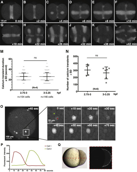
Dynamics of Ca2+ signaling in Tg[βactin2:GCaMP6s]stl351/stl351 embryos at cleavage and blastula stages. (A-L) Still images of GCaMP6s signals during cleavage furrow progression from 2-cell stage to 16-cell stage. (M) Quantification of Ca2+ transient duration before and after MBT. Error bars represent standard deviation; N=4 embryos. ns, not significant. (N) Comparison of Ca2+ transient numbers before and after MBT; N=6 embryos. (O-P) Still images and traces of Ca2+ transients in a time-lapse series at blastula stages. (Q) Time-lapse overlay of GCaMP6s signal from 3.7 hpf to 4 hpf from a single z-section in lateral view. |
Reprinted from Developmental Biology, 430(2), Chen, J., Xia, L., Bruchas, M.R., Solnica-Krezel, L., Imaging early embryonic calcium activity with GCaMP6s transgenic zebrafish, 385-396, Copyright (2017) with permission from Elsevier. Full text @ Dev. Biol.