
Fig. 6
- ID
- ZDB-FIG-180206-28
- Publication
- Bower et al., 2017 - SERCA directs cell migration and branching across species and germ layers
- Other Figures
- All Figure Page
- Back to All Figure Page
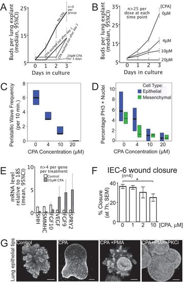
SERCA function controls the onset and rate of lung branching. (A) SERCA function dictates the onset of new buds. Plot of lung bud count versus time in culture for E13 rat lung explants shows the budding rate in controls (no CPA), lack of budding with 20 μM CPA, and resumption of budding when CPA is removed. (B) SERCA function titrates budding rate. Bud count plotted against days in culture. The normal accretion of buds is shown in the absence of CPA (0 μM). Escalating the CPA dose controls the budding rate. At 20 μM, branching is arrested. (C) The frequency of airway peristaltic waves decreases with escalating CPA dose, with statistical significance between each treatment group (P<0.05, Mann-Whitney U test). Median and interquartile range (IQR) are plotted, n>10 for each treatment. (D) Proliferation of lung epithelial and mesenchymal cells decreases with escalating CPA dose, with statistical significance between treatments for each cell type, except 10 µM and 20 µM are equivalent (P<0.05, Mann-Whitney U test). Median and IQR of PH3 positive nuclei are plotted, n>24 for each treatment. (E) SERCA blockade is associated with downregulation of lung morphogens SHH, FGF10, and SMMHC (smooth muscle myosin heavy chain), and significant upregulation of SPRY2, FGF9 and VEGF (qRT-PCR). Error bars indicate 95% confidence interval. (F) SERCA inhibition impairs epithelial cell migration. Plot of percentage closure at 7 h (mean±s.e.m.) of a standardized wound in a confluent monolayer of IEC-6 intestinal epithelial cells treated with 0, 1, 2, or 10 μM CPA. Wound closure is significantly reduced by 10 μM CPA (*P<0.05, one-way ANOVA and Bonferroni multiple comparisons test). (G) Epithelial SERCA blockade halts budding and is rescued by PKC activation. Epithelial tips isolated from E12.5 murine lungs bud in Matrigel with FGF10. Control epithelial tips bud extensively (left panel). 10 μM CPA abolishes budding, despite co-incubation with FGF10 (2nd panel). Budding is rescued by co-treatment with PKC activator (100 nM PMA) (3rd panel). Budding is re-inhibited by PKC inhibition (2.22 μM Bisindolylmaleimide I Hydrochloride), demonstrating that PMA rescue is mediated by PKC (right panel). Scale bars: 100 μm |