
FIGURE
Fig. 2
- ID
- ZDB-FIG-180117-27
- Publication
- Sun et al., 2017 - Anti-inflammatory properties of extracts from Chimonanthus nitens Oliv. leaf
- Other Figures
- All Figure Page
- Back to All Figure Page
Fig. 2
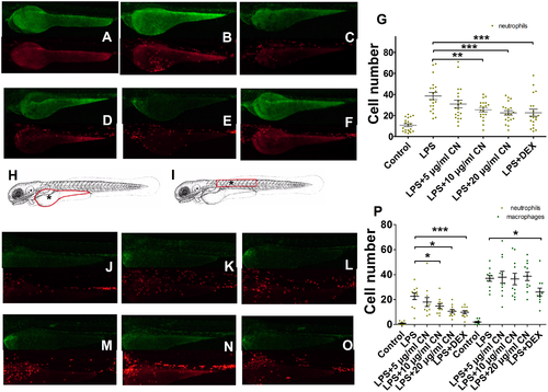
Macrophages and neutrophils migrated to the injured area were count. A-F, images show the macrophages and neutrophils migrated to the yolk microinjected by LPS at 6 hpi. G, neutrophils count in yolk at 6 hpi (n = 20). H and I, microinjection site were marked by * and cell count area were marked by red circle. J-O, images show the macrophages and neutrophils migrated to the somite muscle microinjected by LPS at 6 hpi. P, macrophages and neutrophils count in somite muscle at 6 hpi (n = 11). Data were shown as mean ± S.E. For G and P, * indicates p<0.05, ** is p<0.01, *** is p<0.001. |
Expression Data
Expression Detail
Antibody Labeling
Phenotype Data
Phenotype Detail
Acknowledgments
This image is the copyrighted work of the attributed author or publisher, and
ZFIN has permission only to display this image to its users.
Additional permissions should be obtained from the applicable author or publisher of the image.
Full text @ PLoS One