
FIGURE
Fig. S2
Fig. S2
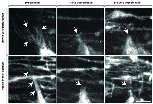
Axotomy of navigating pioneer axon growth cones. Zoomed images of axotomized Tg(sox10:mrfp) growth cones showing pre-axotomy, 1 hour after axotomy and 24 hours after axotomy. The filopodia-like projections that are typical of a growth cone are absent after axotomy. Note the lack of debris from RFP+ glia. Arrows denote growth cone filopodia extensions that are missing after axotomy. Scale bar, 25 μm. |
Expression Data
Expression Detail
Antibody Labeling
Phenotype Data
Phenotype Detail
Acknowledgments
This image is the copyrighted work of the attributed author or publisher, and
ZFIN has permission only to display this image to its users.
Additional permissions should be obtained from the applicable author or publisher of the image.
Full text @ PLoS Genet.