
Fig. 7
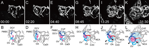
Integration of the hyaloid and ciliary vascular systems (rostral-lateral view). Selected time-lapse images of a living Tg(flk1:EGFP)k7 embryo from 1.25 dpf (S5 Movie) (A, C, E, G, I, and K) and their schematic diagrams (B, D, F, H, J, and L). The time (hours:minutes) from the first frame is labeled in each image (A, C, E, G, I, and K). Rostral is facing right and dorsal is facing upward. The formation of the right ocular vasculature, including the lateral transfer of the OV and the integration of the two systems via the IOC, were observed. Ocular vessels in the schematic diagrams are colored (NAC: red, DCV: blue, OA: pink, OV: sky blue, and CVP: light blue). Arrow in F indicates the connecting portion of the NCA in formation and the CrDI. Arrowheads in J and L indicate the forming IOC, which connects the hyaloid vascular system with the ciliary vascular system. |