
Fig. S7
- ID
- ZDB-FIG-180103-58
- Publication
- Emfinger et al., 2017 - Expression and function of ATP-dependent potassium channels in zebrafish islet β-cells
- Other Figures
- All Figure Page
- Back to All Figure Page
|
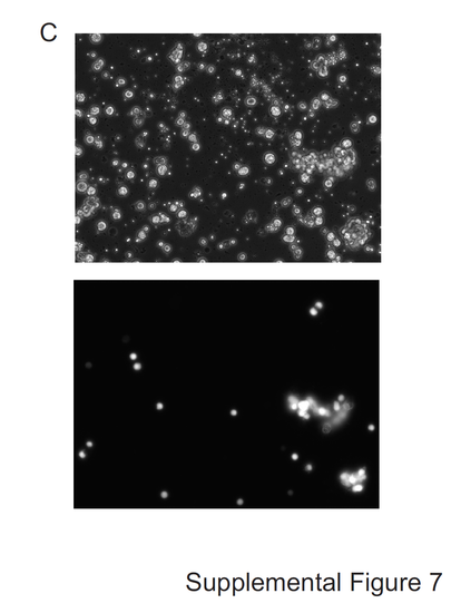
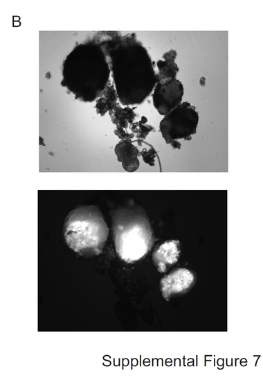
Original microscopy images in Figure 1. (A) Bright field (top) and green fluorescence (bottom) of an adult (12 week old) zebrafish anesthetized on its left side (10x). These images were combined and rotated prior to their use in Figure 1, as indicated in Figure 1 legend. Bright-field image was contrast-enhanced prior to being super-imposed with fluorescent image to improve visibility within final combined image. (B) Bright field (top) and green fluorescence (bottom) images of isolated zebrafish islets (20x). (C) Bright field (top) and green fluorescence (bottom) images of dispersed zebrafish β-cells (40x). (D) Co-localization of insulin and eGFP staining in zebrafish histological sections confirms specificity of insulin promoter driving eGFP (20x). 

|
| Gene: | |
|---|---|
| Fish: | |
| Anatomical Term: | |
| Stage: | Adult |