
Fig. 2
- ID
- ZDB-FIG-171229-2
- Publication
- Shainer et al., 2017 - Novel hypophysiotropic AgRP2 neurons and pineal cells revealed by BAC transgenesis in zebrafish
- Other Figures
- All Figure Page
- Back to All Figure Page
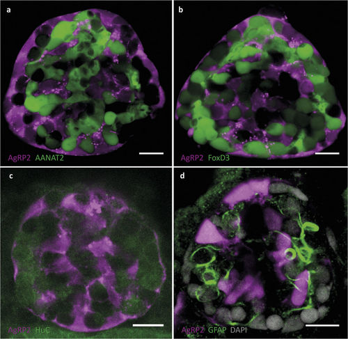
AgRP2 is expressed in uncharacterized pineal cells. (a) Pineal gland of a 5-dpf agrp2:mCherry, Tg(aanat2:EGFP)y9 larva. AgRP2 cells (magenta) do not co-localize with pineal photoreceptor cells (green). (b) Pineal gland of a 5-dpf agrp2:mCherry, Tg(foxd3:EGFP)zf104 larva. AgRP2 cells (magenta) do not co-localize with FoxD3 pineal neurons (green). (c) Double immunostaining of a 5-dpf agrp2:mCherry larva with an antibody against HuC (a neuronal marker) and with anti-RFP. AgRP2 cells (magenta) do not co-localize with HuC-positive cells (green). (d) Immunostaining of a 5-dpf agrp2:EGFP larva with an antibody against GFAP (a marker for pineal interstitial cells) and with anti-EGFP. AgRP2 cells (magenta) do not co-localize with pineal interstitial cells (green). Scale bar, 10 μm. |
| Genes: | |
|---|---|
| Antibody: | |
| Fish: | |
| Anatomical Terms: | |
| Stage: | Day 5 |