
Fig. 3
- ID
- ZDB-FIG-171206-71
- Publication
- Sheets, 2017 - Excessive activation of ionotropic glutamate receptors induces apoptotic hair-cell death independent of afferent and efferent innervation
- Other Figures
- All Figure Page
- Back to All Figure Page
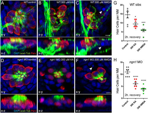
KA or NMDA exposure initiates hair-cell loss in ngn11 morphants comparable to WT siblings. (A–F) Representative max intensity top-down (x-y) and side-view (x-z) projections of imunolabeled Parvalbumin (red), acetylated Tubulin (green), and DAPI (blue) in posterior LL NM7 of 5 dpf WT (A–C) or ngn1 MO (D–F) larvae. Larvae were exposed to DMSO carrier alone (Control) or iGluR agonists for 1 hour, then rinsed and allowed to recover for 2 hours. Exposure to 300 μM KA (B,E) or 300 μM NMDA (C,F) leads to hair-cell loss in both WT and morphants. White arrowheads indicate pyknotic nuclei. Scale bar: 3 μm (G,H) The number of intact hair cells per NM in 5 dpf larvae exposed to DMSO alone, 300 μM KA, or 300 μM NMDA. Each circle represents NM2 or NM7 in an individual larva. There were significantly fewer HCs per NM in KA and NMDA-treated larvae compared to control 2 h. after exposure in both WT siblings (G) and ngn1 (H) MO larvae (*p < 0.05; ***p < 0.001; ****p < 0.0001 defined by Dunnett’s multiple comparisons test). |
| Fish: | |
|---|---|
| Conditions: | |
| Knockdown Reagent: | |
| Observed In: | |
| Stage: | Day 5 |