
Fig. 1
- ID
- ZDB-FIG-171127-57
- Publication
- Takita et al., 2016 - Effects of NDRG1 family proteins on photoreceptor outer segment morphology in zebrafish
- Other Figures
- All Figure Page
- Back to All Figure Page
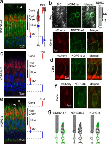
Subcellular localization of NDRG1 family proteins in zebrafish photoreceptors. Adult zebrafish retinas were immunostained with specific anti-NDRG1a-1 (a,b), anti-NDRG1a-2 (c,d) or anti-NDRG1b (e,f) antiserum (all with green signals). Mitochondria and nucleus were counterstained with anti-Tom20 antibody (red signals) and Hoechst 33342 (blue signals), respectively (a,c,e). In a, c and e, approximate positions of ellipsoid containing mitochondria (red bars) and nucleus (blue bars) for each cone type (red/green-, blue- and UV-sensitive cones) and for rods are indicated. NDRG1a-1 was found to be expressed in the ellipsoid but not in the OS in rods (arrowhead in a, and upper panels in b). NDRG1a-1 was also found both in the OS and ellipsoid in cones (a and lower panels in b). NDRG1a-2 was found in the thin process of a cone (c,d). NDRG1b was found in the entire region of a cone (e,f) but not in rod ellipsoid (arrowheads in e). In a, c and e, wildtype zebrafish retinas were used, and in the detailed study in b, d and f, the retinas consisting of cones expressing mCherry-HrasCAAX were used to readily identify cones. (g) Schematic representation of localization of NDRG1 family proteins in adult zebrafish photoreceptors (shown in green). DIC, differential interference contrast image. |
| Antibodies: | |
|---|---|
| Fish: | |
| Anatomical Terms: | |
| Stage: | Adult |