
FIGURE
Fig. 4
- ID
- ZDB-FIG-171127-36
- Publication
- Naylor et al., 2016 - Caudal migration and proliferation of renal progenitors regulates early nephron segment size in zebrafish
- Other Figures
- All Figure Page
- Back to All Figure Page
Fig. 4
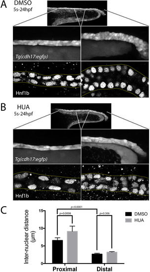
Live morphology of the pronephric tubule. (A,B) Top panels show a lateral view of the pronephric tubule in live Tg(cdh17:egfp) embryos at 24 hpf treated with either DMSO (A) or HUA (B). The two panels below show the same embryos with higher magnification views of the proximal (left) and distal (right) tubule. The bottom panels are single plane lateral views of Hnf1b stained pronephric nuclei in the proximal (left) and distal (right) tubule. (C) Histogram representing the inter-nuclear distances observed in the proximal and distal tubules of DMSO (control) and HUA treated embryos. |
Expression Data
Expression Detail
Antibody Labeling
Phenotype Data
Phenotype Detail
Acknowledgments
This image is the copyrighted work of the attributed author or publisher, and
ZFIN has permission only to display this image to its users.
Additional permissions should be obtained from the applicable author or publisher of the image.
Full text @ Sci. Rep.