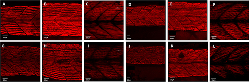
Disruption of myofibril organization during paralysis is reversed by restoring movement in developing skeletal muscle. Control embryos (Embryo Medium alone) and treated embryos (Embryo Medium with Tricaine) were incubated for 7 h starting at 17 hpf. At 24 hpf embryos were removed and control (A) and treated embryos (G) were fixed and stained immediately or put into recovery (Embryo Medium without Tricaine) and (B) control and (H) recovered embryos fixed and stained at 42 hpf. Some embryos were kept in recovery from 24 hpf up to 5 dpf before fixation and staining of both control (C) and recovered larvae (I). Relaxed immotile mutants were fixed and stained at 24 hpf (J), at 42 hpf (K), and 5 dpf (L) as well as motile control siblings at 24 hpf (D), 42 hpf (E) and 5 dpf (F). Anti-myosin antibody (F59) in (A,B,D,E,G,H,J,K), and Rhodamine phalloidin actin labeling in (C,I,F,L) reveals slow muscle fibers. For consistency, the somites imaged were taken at the level where the yolk sac and the yolk sac extension join. Scale bars corresponds to 20 μm.
|
