
Fig. 4
- ID
- ZDB-FIG-171027-34
- Publication
- Grone et al., 2015 - Divergent evolution of two corticotropin-releasing hormone (CRH) genes in teleost fishes
- Other Figures
- All Figure Page
- Back to All Figure Page
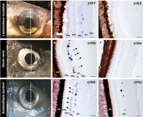
Localization of crh forms in the retina of fishes. Photographs of the eye of each species and adjacent 20 μm transverse sections of the retina reacted with each crh anti-sense probe are shown. (A–C) The spotted gar Lepisosteus oculatus shows crh1 expression in the inner nuclear layer, but crh2 is absent from the retina. (D–F) The zebrafish Danio rerio expresses crhb in the amacrine cell region of the inner nuclear and in the ganglion cell layer (arrows), but crha is absent from the retina. (G–I) The African cichlid fish Astatotilapia burtoni shows crhb label in two different cell types within the inner nuclear layer, amacrine and bipolar cells (arrows). crha was also found in the A. burtoni retina, but was restricted to the region of amacrine cells in the inner nuclear layer. GCL, ganglion cell layer; INL, inner nuclear layer; IPL, inner plexiform layer; OPL, outer plexiform layer; PHL, photoreceptor layer; PE, pigmented epithelium. Lines on (A,D,G) indicate the approximate position of the sections. Scale bars = 1 mm (A,D,G); 25 μm (B,C,E,F,H,I). |
| Genes: | |
|---|---|
| Fish: | |
| Anatomical Terms: | |
| Stage: | Adult |