
Fig. 5
- ID
- ZDB-FIG-170914-28
- Publication
- Scott et al., 2017 - Nuclear/cytoplasmic transport defects in BBS6 underlie congenital heart disease through perturbation of a chromatin remodeling protein
- Other Figures
- All Figure Page
- Back to All Figure Page
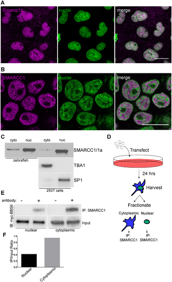
BBS6 and SMARCC1 protein localizations overlap in subcellular compartments. A single z-slice of a confocal stack of cells in a 70% epiboly staged zebrafish embryos stained with a SMARCC1 antibody, nuclei counterstained with TO-PRO-3 (A). SMARCC1 immunostaining of 293T cells shows predominant nuclear localization, nuclei counter-stained with TO-PRO-3 (B). Cellular fractionation of 24hpf zebrafish as well as 293T cells show cytoplasmic and nuclear localization of endogenous Smarcc1a/SMARCC1, TBA1 and SP1 served as cytoplasmic and nuclear fraction controls (C). Experimental design of cellular transfection and fractionation followed by IP on individual sub-cellular fractions (D). Western blot showing myc-BBS6 can coIP with SMARCC1 in the cytoplasm and nucleus (E). Normalizing IP band intensity to input shows stronger interaction in the cytoplasm (F). Scale bars = 20μm. |