
Fig. S1
- ID
- ZDB-FIG-170606-16
- Publication
- Lewis et al., 2017 - Cos2/Kif7 and OSM-3/Kif17 Regulate Onset of Outer Segment Development in Zebrafish Photoreceptors Through Distinct Mechanisms
- Other Figures
- All Figure Page
- Back to All Figure Page
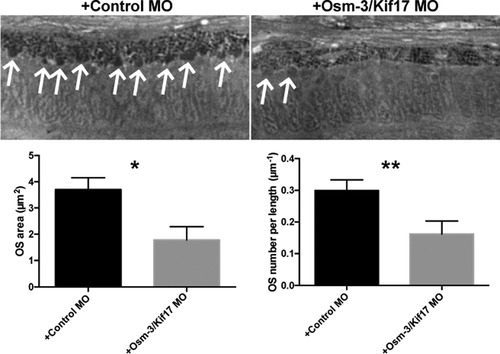
osm-3/kif17 morphants have a reduction in both OS number and size. (Top) Wild-type embryos were injected with either a control morpholino (+Control MO) or osm-3/kif17 splice site junction morpholino (+Osm-3/Kif17 MO) and raised to 72hpf. Plastic histology reveals several evenly sized and uniformly distributed OS in the central retina of control morpholino-injected embryos (arrows), but OS are unevenly distributed and variable in size in osm-3/kif17 morpholino-injected embryos (arrows). (Bottom) Five biological replicates for each genotype were quantified as described above for the average OS area and density. osm-3/kif17 morpholino-injected embryos have approximately half as many OS, and OS that are approximately half the size of controls. |
| Fish: | |
|---|---|
| Knockdown Reagent: | |
| Observed In: | |
| Stage: | Protruding-mouth |
Reprinted from Developmental Biology, 425(2), Lewis, T.R., Kundinger, S.R., Pavlovich, A.L., Bostrom, J.R., Link, B.A., Besharse, J.C., Cos2/Kif7 and OSM-3/Kif17 Regulate Onset of Outer Segment Development in Zebrafish Photoreceptors Through Distinct Mechanisms, 176-190, Copyright (2017) with permission from Elsevier. Full text @ Dev. Biol.