
Fig. 1
- ID
- ZDB-FIG-170606-10
- Publication
- Lagos et al., 2017 - Characterization and Vaccine Potential of Membrane Vesicles Produced by Francisella noatunensis subsp. orientalis in an Adult Zebrafish Model
- Other Figures
- All Figure Page
- Back to All Figure Page
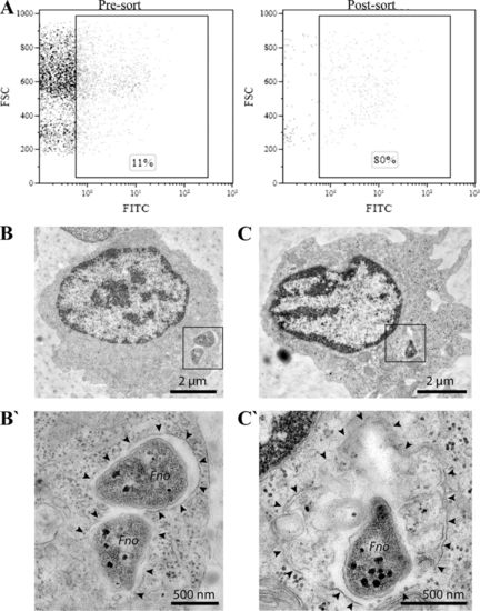
F. noatunensis subsp. orientalis uptake by zebrafish leukocytes in vitro. (A) Representative dot plot before (Pre-sort) and after (Post-sort) cell sorting of leukocytes harvested from adult zebrafish kidney incubated for 1 h with Fno-FITC. Bacterial uptake was quantified by flow cytometry as the percentage of cells with increased FITC fluorescence. (B to C′) Ultrastructural TEM analysis of zebrafish kidney leukocytes with internalized F. noatunensis subsp. orientalis (Fno) after 1 h of incubation. In macrophages (B and C), bacteria reside mostly in tight phagosomes (B′) and only rarely in larger phagolysosomes (C′) with additional luminal cargo. Black arrowheads indicate phagosomal/phagolysosomal membrane. Bars, 2 μm (B to C) and 500 nm (B′ to C′). |